pISSN : 3058-423X eISSN: 3058-4302
Open Access, Peer-reviewed

pISSN : 3058-423X eISSN: 3058-4302
Open Access, Peer-reviewed

Dong Hyun Shim,Jae Hyeong Seo,Hoon Choi
10.17966/JMI.2026.31.3.105 Epub 2026 October 01
Abstract
Recalcitrant onychomycosis—defined as persistent or recurrent, mycologically confirmed onychomycosis despite adequate standard systemic antifungal therapy, including at least one appropriately administered repeat treatment course—represents a multifactorial clinical challenge. Rather than attributing failure to pharmacologic inadequacy alone, this review reframes recalcitrance as a biologically driven phenomenon in which fungal reservoirs may become tolerant to conventional antifungal exposure. The underlying pathophysiology can be categorized into five interacting domains: diagnostic inaccuracy, arthroconidial dormancy, dermatophyte biofilm (dermatophytoma) formation, host immune anergy, and environmental reinfection sources. We have reviewed evidence-based strategies for overcoming recalcitrance, including confirmatory re-diagnosis before retreatment, extended and pulse medication regimens exploiting arthroconidial activation-kill cycles, boosted antifungal protocols (boosted antifungal topical treatment and boosted oral antifungal treatment), biofilm-disrupting and keratolytic adjuncts, laser-assisted drug delivery, and combined systemic and topical therapy. We emphasize environmental decontamination—footwear disinfection, laundry management, and family co-treatment—alongside long-term post-cure prophylaxis, as being essential for preventing relapse and reinfection. Recalcitrant onychomycosis requires a paradigm shift toward laboratory-directed diagnosis and individualized, multimodal treatment beyond the nail itself to achieve a durable cure.
Keywords
Antifungal agents Biofilm Drug resistance Fungal Onychomycosis Recurrence
Onychomycosis is a chronic superficial fungal infection of the nail unit that is predominantly caused by dermatophytes (Trichophyton rubrum and T. mentagrophytes), although nondermatophyte molds and yeasts account for a smaller but clinically significant proportion1,2. Onychomycosis may clinically present with nail discoloration, subungual hyperkeratosis, onycholysis, and onychauxis2. It accounts for approximately 30% of all cutaneous fungal infections and 50% of all instances of nail disease, and it substantially impairs quality of life due to the attendant pain, functional limitations, and psychosocial impact3,4.
Although effective systemic antifungal agents are available, treatment failure following standard oral therapy has been reported in 20-50% of patients5. Clinical nail findings that are associated with poorer response to treatment include subungual hyperkeratosis of a larger extent than 2 mm, involvement of more than 50% of the nail plate, nail matrix involvement, dermatophytoma, total dystrophic onycho- mycosis, two feet-one hand syndrome, and slow nail growth6. The recent emergence of terbinafine-resistant dermatophytes, particularly T. indotineae, has drawn attention to antifungal resistance as a cause of treatment failure7. However, anti- fungal resistance only accounts for part of the problem. Diagnostic error, limited drug delivery to the nail, fungal persistence, host factors, and reinfection can also contribute to poor outcomes.
This narrative review synthesizes available clinical, mycol- ogical, and translational evidence to characterize the factors that underlie treatment failure in onychomycosis and to outline management strategies for recalcitrant onychomycosis, with an emphasis on laboratory-directed and patient-centered care.
While the term is widely used, recalcitrant onychomycosis has no standardized definition, and published studies only apply heterogeneous criteria for cure, treatment failure, and recurrence8-10. For the purposes of this review, a recalcitrant case of onychomycosis is defined as one that is persistent or recurrent and with mycologically confirmed onychomycosis in spite of adequate standard systemic antifungal therapy, including at least one appropriately administered repeat treatment course. Mycological confirmation should demon- strate persistent evidence of fungal infection obtained by direct microscopy and/or fungal culture11,12. Recurrent disease may represent either relapse, which is defined as the reemergence of the original infecting organism or strain following mycol- ogical cure, or reinfection, defined as infection by a new organism or strain following mycological cure11. In routine clinical practice, however, it is often difficult to distinguish these entities in the absence of molecular strain typing or comparable genotypic methods.
Treatment failure is usually multifactorial and extends be- yond antifungal resistance; the following sections examine these contributing factors and the corresponding strategies for management.
3.1 Empirical and inaccurate diagnosis
Onychomycosis is often thought to admit of easy clinical diagnosis, but an accurate diagnosis is frequently problematic; a failure to confirm the causative organism in a case can lead to ineffective, costly therapy and unnecessary drug exposure13,14. Reliance on clinical inspection alone also risks misdiagnosis of nonfungal nail disorders—including malignant entities, such as subungual squamous cell carcinoma and melanoma—that closely mimic onychomycosis (Table 1)15.
|
Benign conditions |
Malignant conditions |
|
Nail psoriasis |
Subungual squamous |
|
Nail lichen planus |
Subungual melanoma |
|
Subungual and |
|
|
Paronychia |
|
|
Subungual exostosis |
|
|
Onychomatricoma |
|
|
Yellow nail syndrome |
|
|
Idiopathic or |
|
The diagnostic modalities range from rapid point-of-care tests to laboratory-based methods. Direct potassium hydroxide (KOH) microscopy provides same-visit results, but it suffers from limited specificity and operator dependency. Lateral-flow immunoassays (e.g., DiafactoryTM) enable dermatophyte antigen detection within as little as 5-10 minutes with improved specificity. Fungal culture remains the reference standard for species identification and antifungal susceptibility profiling, although it requires 1-4 weeks to yield results. The periodic acid–Schiff (PAS)-stained histopathology of nail clip- pings enhances diagnostic accuracy, in particular when the test results for fungal culture have been negative, while polymerase chain reaction (PCR)-based methods provide the greatest sensitivity and specificity, additionally enabling the detection of resistance-associated mutations. Dermoscopy, while it is not mycologically confirmatory, serves as a useful noninvasive adjunct to differentiate onychomycosis from traumatic onycholysis as well as from melanonychia14,15.
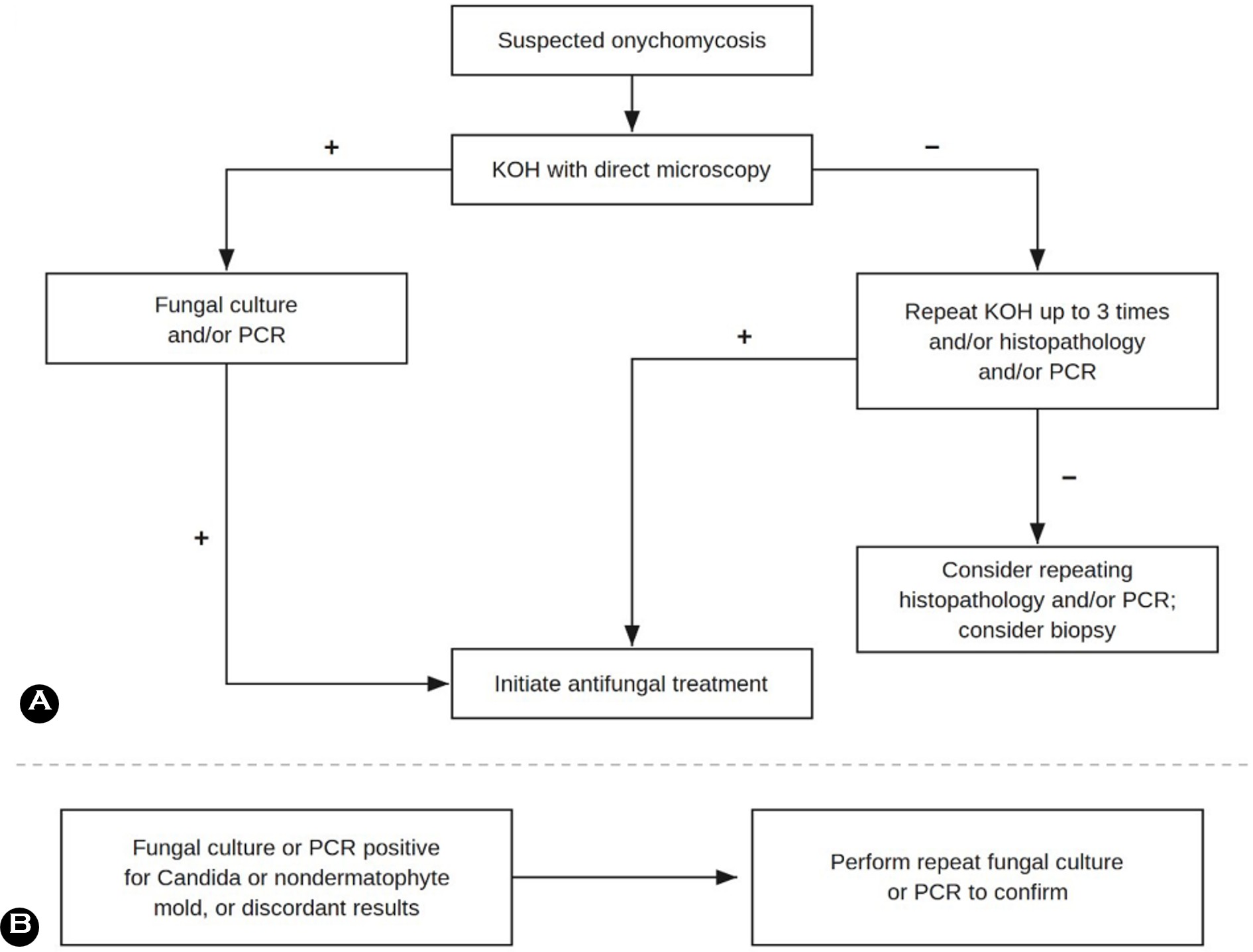
Diagnostic accuracy is influenced by several overlapping challenges. Inadequate specimen collection—in particular from the proximal nail plate rather than the leading edge of infection—markedly reduces sensitivity. Etiological diversity further complicates the decision-making: dermatophytes (predominantly Trichophyton spp.) that account for ~75% of cases in Europe, while nondermatophyte molds, yeasts, and mixed infections make up a significant proportion, requiring species-level identification. Even when applying optimal sam- pling techniques, a discordance between the results of direct microscopy and fungal culture occurs in approximately 33% of cases. A combined diagnostic approach—such as using KOH microscopy with fungal culture or PCR with histopathology—is therefore recommended to maximize diagnostic yield, in particular when nondermatophyte molds or resistant fungal strains are suspected (Fig. 1)15. When mycological diagnosis is delayed, incomplete, or empirically bypassed, the develop- ment of recalcitrant onychomycosis is frequently the clinical consequence.
3.2 Arthroconidia: dormant fungal reservoirs
3.2.1 Formation and biological significance
Arthroconidia, or asexual spores formed via the segmen- tation of fungal hyphae, serve as the principal mechanism of dermatophyte invasion of the nail apparatus and estab- lishment of persistent infection. These propagules are highly prevalent in moist cutaneous environments and nail tissue, forming the primary reservoir for clinical relapse and post- treatment recurrence16. Their pathogenic significance in recalcitrant onychomycosis stems from two critical character- istics: a thickened cell wall conferring increased tolerance to antifungal exposure, and the ability to enter a metabolically quiescent state, rendering the pathogen refractory to fungi- cidal agents that depend on active cellular proliferation17,18.
3.2.2 Environmental triggers of arthroconidial transition
Arthroconidia formation is induced by the hyphal pene- tration of the nail and is accelerated by shifts in the host-tissue microenvironment. In particular, the hypoxia and hyper- capnia resulting from subungual hyperkeratosis, which is a defining characteristic of established onychomycosis, provide an optimal environment for sporulation16,19. T. rubrum readily forms arthroconidia both in vitro and in vivo under these conditions, producing a self-perpetuating cycle of chronic infection16,17.
3.2.3 Implications for antifungal drug efficacy
The clinical impact of arthroconidial dormancy is profound. In an in vitro human nail model inoculated with T. rubrum, the minimum fungicidal concentration (MFC) of terbinafine was approximately 100-fold higher than that determined using standard susceptibility testing20. In a separate study, dormant dermatophytes showed a markedly reduced sus- ceptibility to terbinafine; the complete killing of dormant T. mentagrophytes required 2.0 μg/mL terbinafine, compared with 0.002 μg/mL during the proliferative phase, producing a 1,000-fold difference18. Despite estimated nail terbinafine concentrations exceeding the reported dermatophyte MFC by more than 10-fold, fungal DNA remained detectable by quantitative PCR, with the mean DNA level decreasing to 36% of the baseline by week 1621. These findings provide a plausible mechanistic explanation for treatment failure in cases of recurrence, despite adequate systemic drug exposure and support therapeutic strategies implemented to overcome the protective nail environment and persistent dormant fungal forms.
3.3 Fungal biofilm (dermatophytoma)
3.3.1 Structure and formation
Biofilms are organized, multicellular microbial communi- ties that are encased within a self-produced extracellular matrix (ECM). Unlike planktonic (free-floating) cells, biofilm-embedded organisms have significantly altered gene ex- pression, cooperative metabolic behavior, and substantially increased tolerance to antifungal agents and immune sur- veillance by the host. T. rubrum and T. mentagrophytes initiate biofilm formation within 3 hours of surface contact, and mature biofilms are established by 72 hours; the fungal elements dispersed from the biofilms exhibit higher cytotoxicity than their planktonic counterparts, contributing to local tissue damage and chronicity22.
The in vivo manifestation of a dermatophytic biofilm is the dermatophytoma: a dense, focal accumulation of hyphae, arthroconidia, and keratin in the nail plate, which was first described by Roberts and Evans in 1998. It presents as opaque yellow, orange, and white patches or longitudinal streaks lying along the lateral nail plate, showing poor adherence to the underlying nail bed. Dermatophytomas are associated with a reduced response to standard oral antifungal therapy and are commonly excluded from most randomized clinical trials23.
3.3.2 Determinants of biofilm formation in onychomycosis
Biofilm-producing organisms have been identified in 51.8% of onychomycosis culture samples. Biofilm formation is consistently associated with chronic exposure to humid environments, prolonged prior oral antifungal therapy with- out achieving mycologic cure (≥5 months; 78.5% of biofilm-positive isolates), and recurrent minor nail trauma (72% of cases, p = 0.006). Diabetes mellitus is a major associated comorbidity (53.5% of biofilm-positive cases), likely owing to impaired immune surveillance, and compromised foot hygiene that are secondary to peripheral neuropathy24.
3.4 Host factors
3.4.1 Age, sex, and genetic predisposition
Advanced age continues to be the most consistently iden- tified host risk factor for onychomycosis and its recalcitrant variants, showing a prevalence of 18.2% in patients aged 60-79 years compared with 0.7% in those under 19 years25. This susceptibility is largely driven by age-related immuno- senescence, characterized by a shift toward Th2-skewed immune responses and a reduced fungicidal capacity of phagocytes, facilitating chronic fungal colonization19. Further, the male sex is associated with an approximately threefold higher risk of infection. From a genetic perspective, rare autosomal recessive mutations in CARD9—a key adaptor protein in innate antifungal immunity—have been implicated in severe, disseminated dermatophyte infections, highlighting the role of genetic immune defects in extreme cases25.
3.4.2 Comorbidities and immunosuppression
Diabetes mellitus is a pivotal host risk factor, which operates through hyperglycemia-induced immune dysfunction, periph- eral vascular insufficiency, and diabetic neuropathy, each of which compromises foot hygiene and increases susceptibility to nail trauma. Onychomycosis in patients with diabetes fre- quently serves as an early predictor for diabetic foot syndrome and plantar ulceration and requires vigorous and prolonged antifungal management25. Systemic corticosteroid use, human immunodeficiency virus (HIV) infection, hematologic malig- nancies, and solid-organ transplantation also significantly predispose patients to chronic, recalcitrant dermatophytosis through iatrogenic or disease-related immunosuppression26.
3.4.3 Host immune anergy and Th1/Th2 imbalance
A pivotal but underappreciated phenomenon in recalcitrant onychomycosis is antigen-specific host anergy. Patients who have chronic dermatophytosis (duration >5 years) display a specific lack of immune responsiveness to Trichophyton antigen, indicating selective immune tolerance that is induced by persistent fungal exposure27. This chronic infection is associated with defective phagocytosis of fungal hyphae and a 20-30% reduction in free radical and nitric oxide generation by phagocytes. Further, there is a pathological transition from the Th1-dominant profile of acute infection, associated with effective cell-mediated fungicidal activity, to a Th2-skewed cytokine environment, characterized by elevated IgE and IgG4 levels; this pattern is also observed in atopic patients and linked to heightened susceptibility to dermatophyte infection28,29. These findings, therefore, suggest that host immune dysfunction could contribute to chronicity and treat- ment refractoriness in onychomycosis.
3.5 Environmental and behavioral factors
Environmental and behavioral factors contribute to both susceptibility to and recurrence of onychomycosis. Among these, major risk factors include occlusive footwear, exposure to shared wet environments, repetitive nail trauma, tinea pedis, and intrafamilial exposure25. Footwear and textiles can also be persistent fungal reservoirs, potentially facilitating reinfection30-32. Preventive and environmental interventions to address these factors are discussed in Section 5.
3.6 Nonadherence to treatment
Beyond diagnostic, mycological, and host-related deter- minants, nonadherence to prescribed regimens is an inde- pendent risk factor for recalcitrant onychomycosis. It should be noted that adherence varies substantially by treatment modality: topical agents show the lowest adherence of avail- able options, owing to the prolonged course of treatment (up to 48 weeks) and frequent daily application. Adherence is then also shaped by comorbidities, treatment accessibility, cost, and perceived disease burden—asymptomatic patients who are concerned primarily with cosmetic appearance are particularly apt to prematurely discontinue treatment. Struc- tured adherence counseling is integral to managing recalcitrant onychomycosis33.
3.7 Antifungal resistance
Terbinafine targets squalene epoxidase (SQLE), and missense substitutions at the Leu393 and Phe397 hotspots increase minimum inhibitory concentrations (MICs) by orders of magni- tude; targeted gene replacement has shown that Leu393Phe alone is not sufficient for high-level resistance34,35. These variants are no longer confined to South Asia: terbinafine-resistant Trichophyton indotineae has been reported across Asia, Europe and North America7. Moreover, Phe397Leu and Leu393Phe were recently detected directly in Korean toenail specimens (4 of 106 SQLE-amplified T. rubrum)36. Because T. indotineae cannot be morphologically distinguished within the T. mentagrophytes complex, internal transcribed spacer sequencing, SQLE genotyping, and antifungal susceptibility testing, where available, should be considered when an ad- equate terbinafine course fails, when the disease is extensive or recently acquired abroad, or before committing the patient to prolonged therapy7.
4.1 Confirming diagnosis before retreatment
It is essential to obtain accurate mycological diagnosis before initiating primary treatment and for the retreatment of apparently recalcitrant disease. The differential diagnosis for nail dystrophy is broad: approximately 50% of all dystrophic nail presentations are not fungal, and psoriasis, lichen planus, chronic trauma, and onychogryphosis are common mimics15. Failure to achieve a mycological cure should not be reflexively attributed to drug resistance or biofilm presence if misdiag- nosis, poor compliance, ongoing environmental/household reinfection, and infection with a pathogen not covered by the initial antifungal agent administered are excluded30. Repeated mycological sampling—through direct microscopy, fungal culture, and, where available, PCR with molecular resistance profiling—is strongly recommended to guide sub- sequent therapy14.
4.2 Oral antifungal agents: standard and ex- tended regimens
Terbinafine (250 mg/day continuously for 12 weeks or 500 mg/day pulse therapy for 1 week, followed by 3 weeks off for 3 or 4 cycles) continues to be first-line treatment for dermatophyte onychomycosis, with complete cure rates of approximately 38% across pivotal randomized controlled trials (RCTs) 37. Itraconazole (pulse intake at 400 mg/day for 1 week/month for 3-4 months, or continuous 200 mg/day intake for 12 weeks) is preferred for a confirmed non- dermatophyte mold (NDM) or for Candida infection, given its broader spectrum38. Fluconazole (150-300 mg once weekly until clearance) offers favorable nail penetration and retention for up to 6 months following treatment due to its hydro- philic properties and long plasma half-life38,39. In cases of recalcitrant disease, the duration of treatment may need to be individualized and continue beyond standard regimens, while clinical assessment should take into account the pro- longed time required for complete nail regrowth, which may take up to 12-18 months for toenails instead of adopting a fixed endpoint. Although no RCTs have evaluated durations beyond 12 months, continued exposure is expected to pro- mote fungus-free nail outgrowth and progressively reduce residual dormant arthroconidia21,40,41.
However, extended regimens can prolong systemic exposure. During courses of a standard duration, aminotransferase elevations are not frequent in healthy adults and are no more common than at baseline, so routine interval monitoring adds little; testing is better reserved for hepatic comorbidity, prolonged or repeated courses, and symptoms of hepatic injury42,43. Comedications should be reviewed before an agent is chosen: itraconazole inhibits CYP3A4, and it may interact with drugs such as statins and warfarin, whereas terbinafine inhibits CYP2D6, and it may interact with antidepressants and β-blockers44.
4.3 Pulse regimens: mechanistic rationale in recalcitrance
A pulse terbinafine regimen (250 mg/day, 4 weeks on and 4 weeks off for 2 therapy cycles) achieved cure rates that are statistically equivalent to continuous therapy (risk ratio (RR) 0.97; 95% confidence interval (CI) 0.77-1.23; p = 0.82)45. Pulse regimens may offer a theoretical advantage for re- calcitrant disease: the proposed rationale for this is that drug-free intervals could allow dormant arthroconidia to resume metabolically active hyphal growth, potentially increasing susceptibility to subsequent antifungal exposure46.
4.4 Boosted therapies: boosted antifungal topical treatment (BATT) and boosted oral antifungal treatment (BOAT)
4.4.1 BATT
The BATT approach, developed by Piérard et al., deliberately activates dormant arthroconidia to increase their suscepti- bility to topical antifungal therapy47. In a pilot study for this approach, 13 patients refractory to both oral antifungals and amorolfine monotherapy received weekly amorolfine lacquer, combined with a Sabouraud agar fragment that was applied every other day for 24 hours during the first week of each monthly therapy cycle for two cycles; a mycological cure was achieved in 11 of 13 patients (84.6%), with no relapse at 6-month follow-up.
4.4.2 BOAT
The BOAT strategy extends the boosting concept to the systemic domain and combines a pulse regimen of itra- conazole (400 mg/day for 1 week/month for 3 consecutive months) with Sabouraud agar patches secured to the affected nail for 48 h, repeated on three consecutive occasions (6 days total) after each pulse regimen48. In a pilot study of 10 patients using a paired-nail design, mycological cure at 7 months was achieved in 9 of 10 BOAT-treated nails (90%) but only in 6 of 10 contralateral control nails receiving conventional itraconazole pulse therapy (60%).
4.5 Antibiofilm strategies
The rapid biofilm-forming capacity of T. rubrum and T. mentagrophytes is speculated to contribute to the persistence and recurrence characteristic of onychomycosis22.
4.5.1 Chemical and enzymatic ECM disruption
The biofilm ECM can increase antifungal MIC values up to 1,000-fold, making ECM disruption a prerequisite for adequate drug delivery22,49. Enzymatic agents (DNase I, α-amylase, and lyases) and chemical disruptors (lactic acid, chitosan, terpinen-4-ol nanoparticles, and povidone-iodine) have shown antibiofilm activity in vitro49. In experimental fungal biofilm models, DNase I combined with amphotericin B achieved a 1-5 log10 reduction in the biofilm viability of C. albicans; silver and polylactic-acid nanoparticles demonstrated the strongest antibiofilm effects of the nanoformulations, and liposomal amphotericin B remained the only FDA-approved antifungal nanoformulation for clinical use50.
4.5.2 Keratolytic adjuncts: urea and com- bination vehicles
Urea-based preparations have an important adjunctive role in the management of biofilm-associated and thick-nail onychomycosis. A topical application of 40% urea denatures nail keratin, softens the hyperkeratotic nail plate, and facili- tates the penetration and debridement of the drug. A systematic review of six RCTs (n = 407) confirmed the benefit of the application of urea as an adjunct to oral or topical antifungal medications, most commonly combined with 1% bifonazole51. Additionally, composite formulations, such as K101 nail solution (Emtrix/Naloc: urea, propylene glycol, and lactic acid), exert keratolytic, acidifying, and direct fungicidal effects, with in vitro studies revealing cell wall/membrane disruption in T. rubrum and C. albicans at 50% concen- tration52. Hydroxypropyl chitosan-based ciclopirox 8% hydro- lacquer (P-3051; FullcareTM in Korea) enhanced transungual penetration and achieved superior cure rates versus amorol- fine at 48 weeks of treatment (35% vs. 11.7%; p < 0.001)53.
4.5.3 Laser-assisted drug delivery
A fractional CO2 laser creates micro-channels that increase transungual permeability. In an imaging study using healthy and mycotic human nail samples, fractional CO2 laser micro- poration significantly increased the penetration of topically applied fluorescent tracer throughout the nail plate. Relative to untreated nails, laser pretreatment increased mean tracer delivery by approximately 108% in healthy nails and 33% in mycotic nails, showing the greatest enhancement in the superficial nail layers. These findings are direct mechanistic evidence that fractional laser pretreatment can enhance passive transungual delivery54. Furthermore, fractional CO2 laser combined with topical tioconazole produced higher complete clinical improvement than the use of laser alone or topical tioconazole alone (55% vs. 30% vs. 25%) in a randomized three-arm study of 120 patients55. These findings support the clinical benefit of laser-assisted topical antifungal therapy.
4.6 Combination systemic plus topical regimens
The use of a combination of systemic and topical antifungal therapies is supported by randomized studies indicating improved mycological or combined clinical and mycological outcomes relative to systemic monotherapy56,57. For mixed dermatophyte-NDM infections (~10% of cases diagnosed by PCR), terbinafine with a broad-spectrum topical agent provided additive coverage58. Subinhibitory antifungal ex- posure can promote the selection or expression of resistance-associated phenotypes, therefore, adequate dosing should be maintained through treatment59.
4.7 Treatment of special populations
In elderly patients, who form the highest-risk group, treat- ment should be individualized with due consideration of comorbidities, concomitant medications, disease severity, and expected nail growth rate19,60. In patients with diabetes, early diagnosis and effective treatment are particularly important, along with appropriate foot care and surveillance for com- plications61. In immunocompromised hosts (patients with HIV, organ transplant recipients, and those undergoing cancer therapy), the threshold for systemic therapy should be low, prophylactic protocols could be considered after treatment completion, and surveillance can be maintained for rare, disseminated infection25.
5.1 Footwear decontamination
Footwear is a critical fungal reservoir that is frequently overlooked. Dermatophytes can survive within shoe interiors for extended periods, persisting in shed keratinocytes where moisture from sweat facilitates adherence to the surface of the insole and prevents desiccation of the fungal elements62. Heavily contaminated footwear should be discarded or decontaminated30. Reported decontamination modalities in- clude ozone gas disinfection (a fungicidal agent used against T. rubrum and T. mentagrophytes), germicidal ultraviolet C radiation-based shoe sanitizers (with a wavelength range of 200-300 nm), and microwave irradiation (560 W for 30 seconds, approximately 60℃). These methods have been shown to reduce or eradicate dermatophytes in shoe insoles without substantial material damage63. Antifungal products (e.g., terbinafine solution spray) that are applied to footwear interiors may further reduce fungal contamination, and peri- odic reapplication may help reduce the risk of reinfection62.
5.2 Textile and laundry management
Socks and hosiery are underappreciated vectors of trans- mission and have been implicated in up to 10.46% of recur- rent infections64. High-temperature laundering can effectively reduce fungal burden: C. albicans may be eradicated at 30℃ for 10 minutes, although T. rubrum requires 60℃ for 30 minutes. Aspergillus spp. may survive even at 60℃, requiring higher temperatures or prolonged tumble-drying. Contam- inated garments should be laundered separately to prevent cross-contamination, and washing machines that are used for infected laundry may themselves serve as reservoirs if they are not periodically decontaminated. Patients should change into fresh, absorbent socks once or twice daily, and copper-impregnated antimicrobial socks can offer additional protection63.
5.3 Family cotreatment
Intrahousehold transmission is a statistically significant determinant of recurrence65. All members of a household who are diagnosed with tinea pedis, onychomycosis, or plantar scaling should be evaluated and, when appropriate, treated simultaneously with the index patient64. Preventive measures—including antifungal foot powder, avoidance of barefoot contact with shared surfaces, and the use of indi- vidual towels and nail-care implements—should be reinforced within households30,62,64.
5.4 Patient education and lifestyle modification
Adherence to pharmacological treatment and preventive hygiene measures is among the most important modifiable determinants for treatment outcomes. Patient education should emphasize keeping the feet and hands clean and dry, regular nail trimming, rotating and thoroughly drying foot- wear, avoiding shared nail care instruments and towels, using footwear in high-risk communal wet areas, and seeking prompt medical evaluation for new signs of fungal infection. Patient education and preventive counseling are important components for long-term strategies to reduce onychomycosis recurrence63,64,66. A summary of recommended patient educa- tion points and lifestyle modifications for reducing recurrence is presented in Table 2.
|
Category |
Recommendation |
Rationale |
|
Foot and
nail hygiene |
Keep feet clean and thoroughly dry; trim nails |
Reduces moisture and nail-barrier disruption that
facilitate fungal persistence |
|
Footwear
and textiles |
Use well-fitting, breathable footwear; rotate and |
Moist footwear and contaminated textiles fungal
reservoirs |
|
Decontamination
and |
Avoid sharing nail-care tools, towels, slippers, or |
Limits indirect transmission and |
|
Household
and concurrent |
Identify and treat coexisting tinea pedis and |
Untreated skin infection or infected |
|
Early
treatment |
Seek evaluation and initiate appropriate |
Early treatment may limit disease progression and
transmission |
|
Post-treatment
prevention |
Consider prolonged preventive measures, |
Patients with previous onychomycosis |
The chronic and recurrent nature of onychomycosis—related to slow nail regrowth (12-18 months for toenails), per- sistent environmental reservoirs, and host-related susceptibility—supports the consideration of posttreatment preventive strategies28,67,68. Topical antifungal therapy applied twice weekly to previously affected nails and surrounding skin following the completion of curative therapy provides the principal evidence base for this approach. A 10% efina- conazole solution, for example, can keep nail-plate concen- trations above MICs for T. rubrum for up to 2 weeks following the last application, providing a favorable pharmacokinetic profile for intermittent prophylactic use63.
In high-risk groups—including patients with diabetes mel- litus, peripheral vascular disease, or documented household transmission—durations of prophylaxis of 2-3 years or more are recommended, addressing both affected nails and the periungual skin/web spaces as reservoirs of residual tinea pedis63,64.
Recalcitrant onychomycosis refers to more than persistent fungal infection, referring to a multifactorial pathogenic condition involving dormant arthroconidia, protective biofilm matrices, host-related susceptibility, and persistent environ- mental reservoirs. Effective management requires the co- ordinated, multimodal framework that is presented in Table 3, integrating laboratory-confirmed diagnosis, appropriate antifungal selection, improved strategies for drug delivery and to address biofilm-associated persistence, individualized treatment duration, environmental control, and recurrence prevention.
|
Dimension |
Core strategy |
Concrete interventions |
|
1.
Precision diagnosis |
Mycological confirmation and pathogen |
PCR-based species profiling for suspected |
|
2. Targeted
antifungal selection |
Agent selection guided by species identity, |
Individualized oral/topical regimen accounting |
|
3. Biofilm
and |
Co-interventions to overcome dormant |
BATT, BOAT, |
|
4.
Immunologically |
Treatment extended until complete |
Prolonged courses in immunocompromised |
|
5.
Environmental |
Concurrent decontamination of fungal reservoirs and
household co-treatment. |
Structured footwear, textile, and household |
|
6. Long-term
surveillance |
Maintenance therapy and monitoring to |
Maintenance topical prophylaxis protocol with |
|
BATT, boosted antifungal topical treatment; BOAT,
boosted oral antifungal treatment; NDM, nondermatophyte mold; PCR, polymerase
chain reaction. The goal is not simply temporary mycological cure,
but durable disease control and prevention of recurrence across all
modifiable domains of the host–pathogen–environment triad. |
||
The achievement of durable control of recalcitrant onycho- mycosis requires clinicians to look beyond the affected nail in particular by addressing fungal reservoirs, host and environmental factors, treatment adherence and long-term strategies to reduce recurrence.
References
1. 1. Gupta AK, Versteeg SG, Shear NH. Onychomycosis in the 21st Century: An update on diagnosis, epidemiology, and treatment. J Cutan Med Surg 2017;21:525-539 https://dx.doi.org/10.1177/1203475417716362 PMID:28639462
Google Scholar
Congratulatory MessageClick here!