pISSN : 3058-423X eISSN: 3058-4302
Open Access, Peer-reviewed

pISSN : 3058-423X eISSN: 3058-4302
Open Access, Peer-reviewed

Geon-Jong Lee,Hyun-Ji Ryu,Kijae Sung,Kyung-Hwa Nam,Jin Park
10.17966/JMI.2026.31.3.145 Epub 2026 October 01
Abstract
Mycoplasma pneumoniae-induced rash and mucositis (MIRM) is an infection-related mucocutaneous eruption characterized by severe mucositis at multiple sites with minimal or absent cutaneous involvement. Its clinical presentation may mimic Stevens-Johnson syndrome, making accurate differentiation crucial, especially in pediatric patients. A four-year-old boy presented with high fever, conjunctival injection, and extensive erosions with hemorrhagic crusting of the lips and oral mucosa. Approximately three weeks before the onset of mucocutaneous lesions, he had developed upper respiratory symptoms and was treated with multiple antibiotics. Physical examination revealed diffuse oral mucosal erosions with extensive hemorrhagic crusting of the lips and some perianal erosions with crusting. Skin involvement was limited to focal crusted erosions on the forehead and a few crusted papules on the trunk. Polymerase chain reaction detected Mycoplasma pneumoniae, supporting the MIRM diagnosis. He was treated with clarithromycin, intravenous immunoglobulin (1 g/kg/day for two days), intravenous methylprednisolone (initially 2 mg/kg/day, followed by tapering), and empirical cefazolin for a possible secondary bacterial infection. His fever resolved within 24 hours after admission, and his mucocutaneous lesions, including conjunctival involvement, gradually improved. Complete recovery was achieved within one month without scarring or complications. MIRM should be considered in children presenting with predominant multisite mucositis and sparse skin findings following atypical pneumonia to enable appropriate management and avoid inappropriate drug-allergy labeling.
Keywords
Mycoplasma pneumoniae Mycoplasma pneumoniae-induced rash and mucositis Reactive infectious mucocutaneous eruption Stevens-Johnson syndrome
Mycoplasma pneumoniae is a common cause of community-acquired respiratory tract infections in children and can trigger various extrapulmonary manifestations, including distinct mucocutaneous eruptions1. Mycoplasma pneumoniae-induced rash and mucositis (MIRM) is an infection-related clinical entity that is characterized by pro- minent mucositis that typically involves the oral, ocular, and anogenital mucosa with absent or minimal cutaneous in- volvement1,2.
Historically, MIRM was classified within the spectrum of erythema multiforme or Stevens–Johnson syndrome/toxic epidermal necrolysis (SJS/TEN). However, it is now recognized as a distinct clinical entity due to its characteristic mucositis-dominant morphology, limited cutaneous lesions, and gener- ally favorable prognosis1,2. Nevertheless, antibiotics or anti- pyretics are frequently administered during the prodromal respiratory phase before mucocutaneous symptoms manifest, which leads MIRM to often be misdiagnosed as drug-induced SJS/TEN. This paper reports a pediatric case of mucositis-dominant MIRM that closely mimicked SJS and highlights key clinical clues that can be used for accurate differentiation.
A four-year-old boy presented with high fever, bilateral conjunctival injection, painful oral erosions, and severe hemor- rhagic crusting of the lips. Approximately three weeks before the onset of the mucocutaneous lesions, he had developed a cough and intermittent fever, for which he was treated at a local pediatric clinic for a suspected upper respiratory tract infection or pneumonia. During this approximately three-week respiratory prodrome, he received several courses of oral antibiotics for the persistent respiratory symptoms, including cefixime and amoxicillin/clavulanate. Three days before presentation at our center, he developed an acute fever with temperatures up to 40℃, lip fissuring, conjunctival injection, and rapidly progressing oral mucosal lesions. He was initially admitted to a secondary hospital with suspected Kawasaki disease or SJS, where intravenous methylpred- nisolone (1 mg/kg/day) was administered for two days and clarithromycin was initiated. Because he remained febrile and mucosal continued worsening, he was transferred to our institution after the three-day admission.
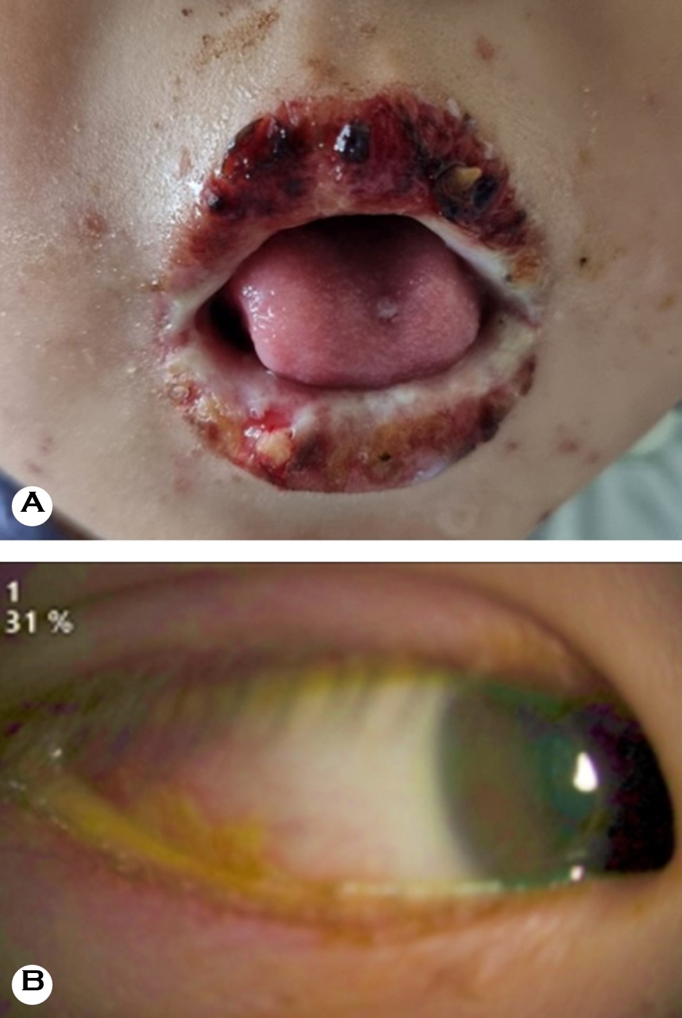
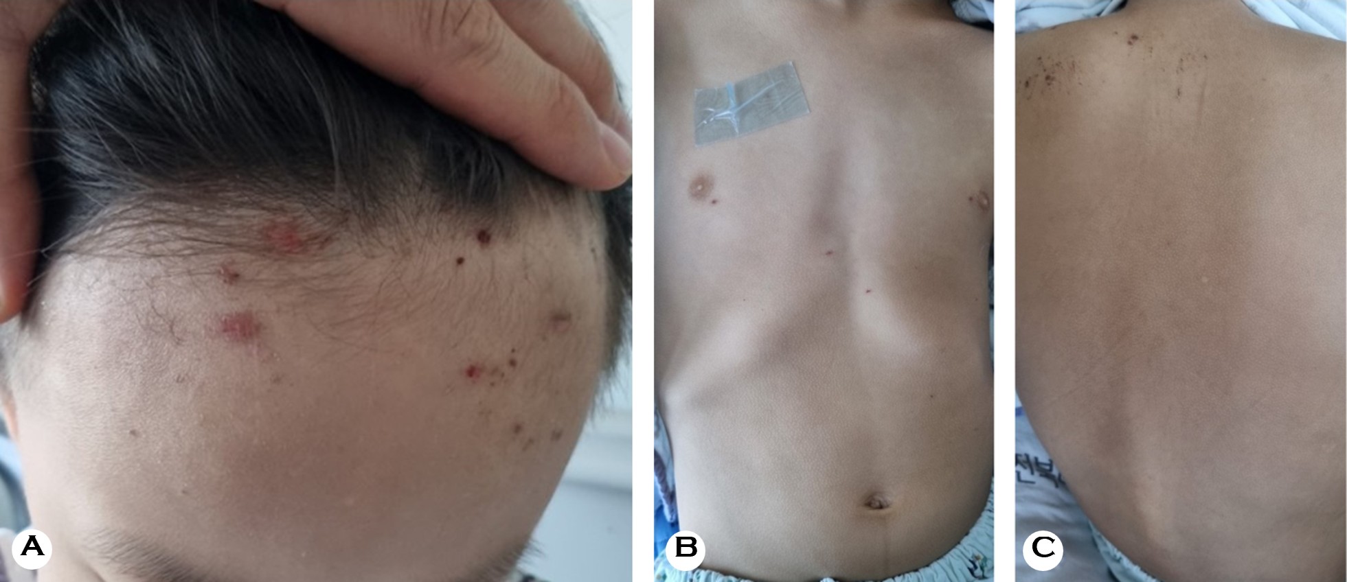
Physical examination upon admission revealed diffuse pain- ful erosions and intense erythema involving the entire oral mucosa and lips, covered with extensive hemorrhagic crusting and accompanied by bilateral conjunctival injection (Figs. 1A and B). Perianal erosions with crusting were also present. Cutaneous involvement was remarkably sparse and restricted to focal crusted erosions on the forehead and a few scattered erythematous crusted papules on the trunk (Figs. 2A-C). There were no generalized bullae, areas of epidermal detachment, or typical targetoid lesions. The Nikolsky sign was negative.
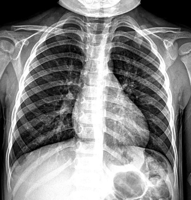
Laboratory findings revealed an elevated erythrocyte sedi- mentation rate of 57 mm/hr and a C-reactive protein level of 51.0 mg/L. Serum amylase was elevated at 765 U/L, but liver and renal function test results were within normal ranges. Serum lipase was also within the normal range (12 U/L). Serum amylase subsequently decreased to 61 U/L before discharge, and follow-up serum lipase remained within the normal range (20 U/L). Abdominal ultrasonography performed during the outpatient follow-up discovered no abnormal findings. Chest radiography demonstrated increased inter- stitial markings in both perihilar regions, which was consistent with bronchopneumonia (Fig. 3). Serologic testing revealed positive IgM and IgG antibodies against M. pneumoniae, and a respiratory multiplex polymerase chain reaction assay detected M. pneumoniae DNA.
Based on the earlier respiratory prodrome, microbiologic evi- dence of M. pneumoniae infection, severe multisite mucositis, sparse cutaneous involvement, and the absence of epidermal detachment, the patient was diagnosed with MIRM. Clarithro- mycin, which had been initiated at the secondary hospital, was continued after his transfer for a total treatment duration of six days. The patient also received intravenous cefazolin for suspected secondary bacterial infection, intravenous immuno- globulin (IVIG; 1 g/kg/day for 2 days), and intravenous methyl- prednisolone was administered at 2 mg/kg/day for two days, then tapered by half every two days for a total of six days. This was administered alongside meticulous supportive mucosal care and urgent ophthalmologic evaluation.
The patient remained febrile at the time of transfer, but the fever subsided within 24 hours after admission to our institution, or approximately three days after initiation of clarithromycin. The respiratory and mucocutaneous symptoms subsequently improved. Laboratory abnormalities, including the inflammatory markers and hyperamylasemia, improved within one week of admission. At the one-month follow-up, all mucocutaneous lesions had completely resolved without scarring, ocular sequelae, or recurrence.
This case demonstrates the importance of recognizing MIRM as a mucositis-dominant eruption that can closely mimic SJS/TEN in pediatric patients. The patient exhibited severe oral and ocular mucositis after a prolonged respiratory prodrome and recent antibiotic exposure, which initially raised concerns about a drug-induced SJS. However, several critical features pointed toward MIRM, including the highly limited cutaneous involvement, the absence of generalized bullae or epidermal detachment, a negative Nikolsky sign, a clear respiratory prodrome, and a laboratory-confirmed M. pneumoniae infection. Kawasaki disease was considered less likely because its other characteristic clinical findings, including extremity changes, were absent.
Canavan et al.'s1 systematic review defined MIRM by its prominent mucosal disease with variable, but usually sparse, skin involvement. Oral, ocular, and urogenital mucositis were reported in 94%, 82%, and 63% of cases, respectively, while cutaneous lesions were absent in 34% and sparse in 47%1. This pattern was consistent with the above patient's presentation, in which severe oral and ocular mucositis with additional perianal involvement predominated and skin involvement remained limited.
Respiratory symptoms generally precede M. pneumoniae-associated mucocutaneous manifestations. In a prospective pediatric cohort by Meyer Sauteur et al.6, three patients with MIRM had prodromal fever and respiratory symptoms for 8-11 days, whereas patients with maculopapular eruptions had prodromal periods of 7-13 days. The approximately three-week respiratory prodrome observed in the above patient was longer than that reported in the MIRM cases in Meyer Sauteur et al.'s6 cohort. However, the timing of an eruption alone does not distinguish MIRM from other M. pneumoniae-associated cutaneous manifestations. The MIRM diagnosis in this patient was supported primarily by the characteristic phenotype of severe multisite mucositis with only sparse cutaneous involvement.
In South Korea, macrolide resistance is an important con- sideration in the treatment of M. pneumoniae pneumonia. A recent multicenter study reported a macrolide resistance rate of 87.0% among Korean children and adolescents with M. pneumoniae pneumonia during the 2023 epidemic7. However, microbiologic macrolide resistance does not neces- sarily correspond to clinical unresponsiveness. Macrolide-unresponsive pneumonia has been defined as a persistent fever of ≥38℃ after ≥72 hours of macrolide therapy. Only 16.3% of hospitalized children met this clinical definition in a separate Korean pediatric cohort8. No specific testing for macrolide resistance was performed in the above patient; therefore, microbiologic macrolide resistance could not be determined. The fever subsided by approximately the third day of clarithromycin therapy, which was followed by improve- ment in his respiratory symptoms. Systemic corticosteroids and IVIG were administered concomitantly, which precluded the attribution of this improvement solely to clarithromycin. Nevertheless, this clinical course did not provide clear evidence of macrolide-unresponsive pneumonia.
Distinguishing MIRM from SJS/TEN has profound practical implications. MIRM carries a significantly more favorable prognosis, with a lower risk of long-term morbidity1,2. Further- more, identifying M. pneumoniae as the likely infectious trigger prevents a mistaken attribution of an eruption to preceding antibiotics, which helps avoid an inappropriate drug-allergy label that unnecessarily restricts a patient's future therapeutic options2.
While the broader term of "reactive infectious muco- cutaneous eruption" has been proposed to encompass similar mucositis-predominant presentations triggered by other pathogens (e.g., Chlamydia pneumoniae)3,4, MIRM remains the most thoroughly characterized prototype. Currently, there are no consensus-based treatment guidelines for MIRM. Supportive care (e.g., hydration, pain control, topical mucosal care, ophthalmologic prevention) remains the cornerstone of management1,2, but systemic corticosteroids and IVIG are often utilized in severe or progressive cases1,2,5. In the above patient, a combination of antimicrobial therapy and immuno- modulatory interventions (IVIG and systemic corticosteroids) was followed by rapid clinical improvement and complete recovery without sequelae. Further studies are needed to clarify the efficacy of these therapeutic interventions in MIRM.
This case highlights the importance of considering MIRM in children presenting with severe multisite mucositis and minimal cutaneous involvement following a respiratory illness. Recognition of this characteristic presentation is important for distinguishing MIRM from SJS/TEN, avoiding inappropriate attribution to preceding medications, and guiding appropriate management and follow-up.
References
1. Canavan TN, Mathes EF, Frieden I, Shinkai K. Mycoplasma pneumoniae-induced rash and mucositis as a syndrome distinct from Stevens-Johnson syndrome and erythema multiforme: A systematic review. J Am Acad Dermatol 2015;72:239-245 https://dx.doi.org/10.1016/j.jaad.2014.06.026
Google Scholar
2. Gandelman JS, Kim EY, Grzegorczyk AM, Zejnullahu K, Edson RS. Mycoplasma pneumoniae-induced rash and mucositis in a previously healthy man: A case report and brief review of the literature. Open Forum Infect Dis 2020;7:ofaa437 https://dx.doi.org/10.1093/ofid/ofaa437
Google Scholar
3. Ramien ML. Reactive infectious mucocutaneous eruption: Mycoplasma pneumoniae-induced rash and mucositis and other parainfectious eruptions. Clin Exp Dermatol 2021;46:420-429 https://dx.doi.org/10.1111/ced.14404
Google Scholar
4. Mayor-Ibarguren A, Feito-Rodriguez M, González-Ramos J, Del Rosal-Rabes T, González-Sainz FJ, Sánchez-Orta A, et al. Mucositis secondary to Chlamydia pneumoniae infection: Expanding the Mycoplasma pneumoniae-induced rash and mucositis concept. Pediatr Dermatol 2017;34:465-472 https://dx.doi.org/10.1111/pde.13140
Google Scholar
5. Chen N, Li M. Case report and literature review: Clinical characteristics of 10 children with Mycoplasma pneumoniae-induced rash and mucositis. Front Pediatr 2022;10:823376 https://dx.doi.org/10.3389/fped.2022.823376
Google Scholar
6. Meyer Sauteur PM, Theiler M, Buettcher M, Seiler M, Weibel L, Berger C. Frequency and clinical presentation of mucocutaneous disease due to Mycoplasma pneu- moniae infection in children with community-acquired pneumonia. JAMA Dermatol 2020;156:144-150 https://dx.doi.org/10.1001/jamadermatol.2019.3602
Google Scholar
7. Lee JK, Lee T, Kim YJ, Kim DR, Shin A, Kang HM, et al. Clinical manifestations, macrolide resistance, and treat- ment utilization trends of Mycoplasma pneumoniae pneumonia in children and adolescents in South Korea. Microorganisms 2024;12:1806 https://dx.doi.org/10.3390/microorganisms12091806, PMID:39338480
Google Scholar
8. Ha SG, Oh KJ, Ko KP, Sun YH, Ryoo E, Tchah H, et al. Therapeutic efficacy and safety of prolonged macrolide, corticosteroid, doxycycline, and levofloxacin against macrolide-unresponsive Mycoplasma pneumoniae pneu- monia in children. J Korean Med Sci 2018;33:e268 https://dx.doi.org/10.3346/jkms.2018.33.e268 PMID:30344461
Google Scholar
Congratulatory MessageClick here!