pISSN : 3058-423X eISSN: 3058-4302
Open Access, Peer-reviewed

pISSN : 3058-423X eISSN: 3058-4302
Open Access, Peer-reviewed

Seowon Song,Subin Lee,Hoon Kang,Jung Eun Kim
10.17966/JMI.2026.31.3.150 Epub 2026 October 01
Abstract
Biologic agents are widely used for severe psoriasis and are generally safe, but infections may develop or worsen during therapy and mimic disease exacerbation. We report a 24-year-old woman with psoriasis treated with guselkumab who developed sudden, widespread worsening of oozing psoriasiform lesions. Skin biopsy revealed dermatophytosis consistent with tinea incognito. Three weeks of oral and topical antifungal therapy resolved the fungal infection, while residual psoriatic lesions continued to improve with guselkumab. Guselkumab has been continued for 1 year without recurrence or reinfection. Although baseline mycological testing was unavailable, the clinical course indicates that a pre-existing, clinically inapparent dermatophyte infection may have become clinically evident and spread extensively during guselkumab therapy. This case highlights the importance of considering fungal infections in patients receiving biologic therapy who develop atypical psoriasis worsening and supports early mycological evaluation when clinically indicated.
Keywords
Guselkumab Interleukin-23 inhibitor Psoriasis Tinea incognito
Psoriasis is a chronic inflammatory skin disease driven by dysregulation of the IL-23/Th17 immune axis. Guselkumab, a monoclonal antibody targeting the interleukin-23 (IL-23) p19 subunit, has demonstrated high efficacy and a favorable safety profile in patients with moderate-to-severe psoriasis1. Although fungal infections are reported less frequently with IL-23 than IL-17 inhibitors, they remain a potential adverse event. Furthermore, fungal infections may present atypically in patients receiving biologic therapies, which can complicate clinical recognition and potentially delay accurate diagnosis and appropriate treatment.
Herein, we report a case of tinea incognito with extensive cutaneous involvement that developed during guselkumab treatment in a patient with psoriasis. This case highlights the importance of considering fungal infections in the differential diagnosis of unexpected or atypical disease worsening of psoriasis during IL-23 inhibitor therapy and underscores the value of early mycological evaluation when clinically indicated.
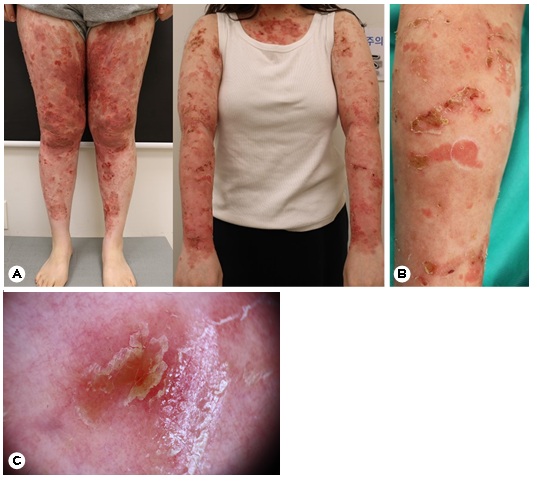
A 24-year-old woman presented with diffuse, painful, and tender erythematous, scaly plaques with yellowish serous exudate and crusting involving nearly the entire body, 11 days after receiving her second injection of guselkumab (Fig. 1). A few targetoid lesions were also noted. The patient had a 10-month history of biopsy-proven severe plaque psoriasis without scalp or nail involvement and no other significant medical history. Despite several months of cyclosporine, methotrexate, and topical betamethasone/calcipotriol, her response was inadequate. She was therefore started on guselkumab while continuing topical treatment. Mild im- provement of the psoriatic lesions was noted after the first injection. However, 3 days after the second injection, the patient developed sudden, widespread worsening of the psoriasiform lesions, with prominent oozing and near-total body involvement.
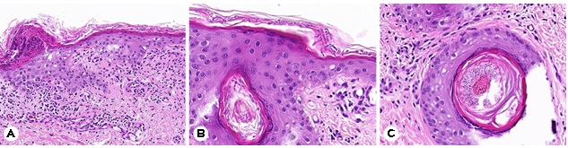
To differentiate psoriatic exacerbation, guselkumab-related adverse effects, and other conditions, a skin biopsy was per- formed. Histopathology revealed numerous fungal hyphae in the stratum corneum and hair follicles, consistent with dermatophytosis (Fig. 2). Culture and molecular methods to identify the specific causative dermatophyte were not performed. Based on the clinical and histopathologic findings, the patient was diagnosed with tinea incognito.
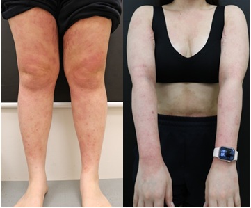
The patient was treated with oral terbinafine, topical amorolfine, and isoconazole for 3 weeks. The fungal com- ponent resolved completely, and the residual psoriatic lesions continued to improve with ongoing guselkumab therapy (Fig. 3). Guselkumab therapy was continued for 1 year, with no evidence of tinea recurrence or reinfection during follow-up.
In the present case, the patient developed sudden, wide- spread worsening of psoriasiform lesions with oozing after the second guselkumab injection, and dermatophytosis was confirmed by skin biopsy, consistent with tinea incognito. The fungal infection resolved completely following antifungal treatment, while the residual psoriatic lesions continued to improve with guselkumab. No tinea recurrence was observed during the subsequent year of guselkumab treatment. Be- cause mycological examination was not performed before guselkumab initiation, it remains unclear whether the der- matophyte infection predated treatment or developed there- after. Nevertheless, the clinical course indicates that a pre-existing, clinically inapparent dermatophyte infection may have become evident and disseminated during guselkumab treatment. Although IL-23 blockade may have contributed through indirect modulation of IL-17-mediated antifungal immune response, a causal relationship between guselkumab and the development or progression of the infection cannot be established.
IL-23 inhibitors appear to be less strongly associated with superficial fungal infections than IL-17 inhibitors. A multi-cohort analysis found a higher risk of candidiasis among patients receiving IL-17 inhibitors than among controls or those treated with IL-23 inhibitors, with no significant difference between IL-23 inhibitor-treated patients and controls2. Although IL-23 inhibitors do not directly inhibit IL-17, IL-23 blockade may indirectly modulate the IL-17-mediated anti- fungal immunity, potentially contributing to fungal infections during IL-23 inhibitor therapy3. Risk factors for fungal in- fections in psoriasis include longer disease duration, higher PASI scores, scalp or nail involvement, older age at biologic therapy initiation, and diabetes mellitus4,5. Our patient was a young woman with a relatively short history and no identifiable risk factors for fungal infection.
Prolonged topical corticosteroid use before and during the early phase of biologic therapy may also have contributed to the development or progression of the superficial fungal infection by suppressing local immunity. Corticosteroid-induced reduction in local inflammation may mask typical clinical features of fungal infection, such as erythema and scaling, causing lesions to resemble psoriasis closely. Moreover, dermatophyte infection may occasionally trigger psoriatic lesions through the Koebner phenomenon, further com- plicating the clinical presentation6. These factors can make it difficult to distinguish tinea incognito from psoriasis based on clinical findings alone. Therefore, mycological evaluation should be considered in patients with psoriasis who develop sudden or atypical worsening during biologic therapy. Early diagnosis may enable prompt antifungal treatment and help avoid unnecessary changes to effective psoriasis therapy.
This case highlights that fungal infection may mimic a psoriasis flare during IL-23 inhibitor therapy and should be considered in the differential diagnosis of treatment failure or disease worsening. Clinicians should maintain a high index of suspicion for fungal infection in patients with sudden or atypical disease worsening during biologic therapy and consider early mycological evaluation to facilitate timely diag- nosis and appropriate management. When a fungal infection is confirmed and effectively treated, an otherwise effective biologic therapy may be continued with appropriate clinical monitoring.
References
1. Reich K, Armstrong AW, Foley P, Song M, Wasfi Y, Randazzo B, et al. Efficacy and safety of guselkumab, an anti-interleukin-23 monoclonal antibody, compared with adalimumab for the treatment of patients with moderate to severe psoriasis with randomized withdrawal and retreatment: Results from the phase III, double-blind, placebo- and active comparator-controlled VOYAGE 2 trial. J Am Acad Dermatol 2017;76:418-431 https://dx.doi.org/10.1016/j.jaad.2016.11.042
Google Scholar
2. Islam RK, Maltese A, Lipner SR. Risk of fungal infection in psoriasis patients treated with IL-17 and IL-23 inhibitors: a multi-cohort study using real-world data. Arch Dermatol Res 2025;317:678 https://dx.doi.org/10.1007/s00403-025-04188-w
Google Scholar
3. Netea MG, van de Veerdonk FL. Anti-interleukin-23 autoantibodies and severe infections. N Engl J Med 2024; 390:1143-1146 https://dx.doi.org/10.1056/NEJMe2400475
Google Scholar
4. Tuerhong T, Nasier M, Maimaiti R, Yan Z, Gang Z, Jinxia H, et al. Pharmacological implications of psoriasis and superficial fungal infections: analysis of risk factors and underlying mechanisms. Pak J Pharm Sci 2025;38:737-743 PMID:40556278
Google Scholar
5. Minami Y, Hiruma J, Harada K, Fujimori K, Suzuki R, Mori M, et al. Risk of fungal infection in patients with psoriasis receiving biologics: a retrospective single-center cohort study. J Am Acad Dermatol 2025;92:108-115 https://dx.doi.org/10.1016/j.jaad.2024.09.037
Google Scholar
6. Zacharopoulou A, Tsiogka A, Tsimpidakis A, Lamia A, Koumaki D, Gregoriou S. Tinea incognito: challenges in diagnosis and management. J Clin Med 2024;13:3267 https://dx.doi.org/10.3390/jcm13113267
Google Scholar
Congratulatory MessageClick here!